Microscopic connections in Natural Light
Work in Progress
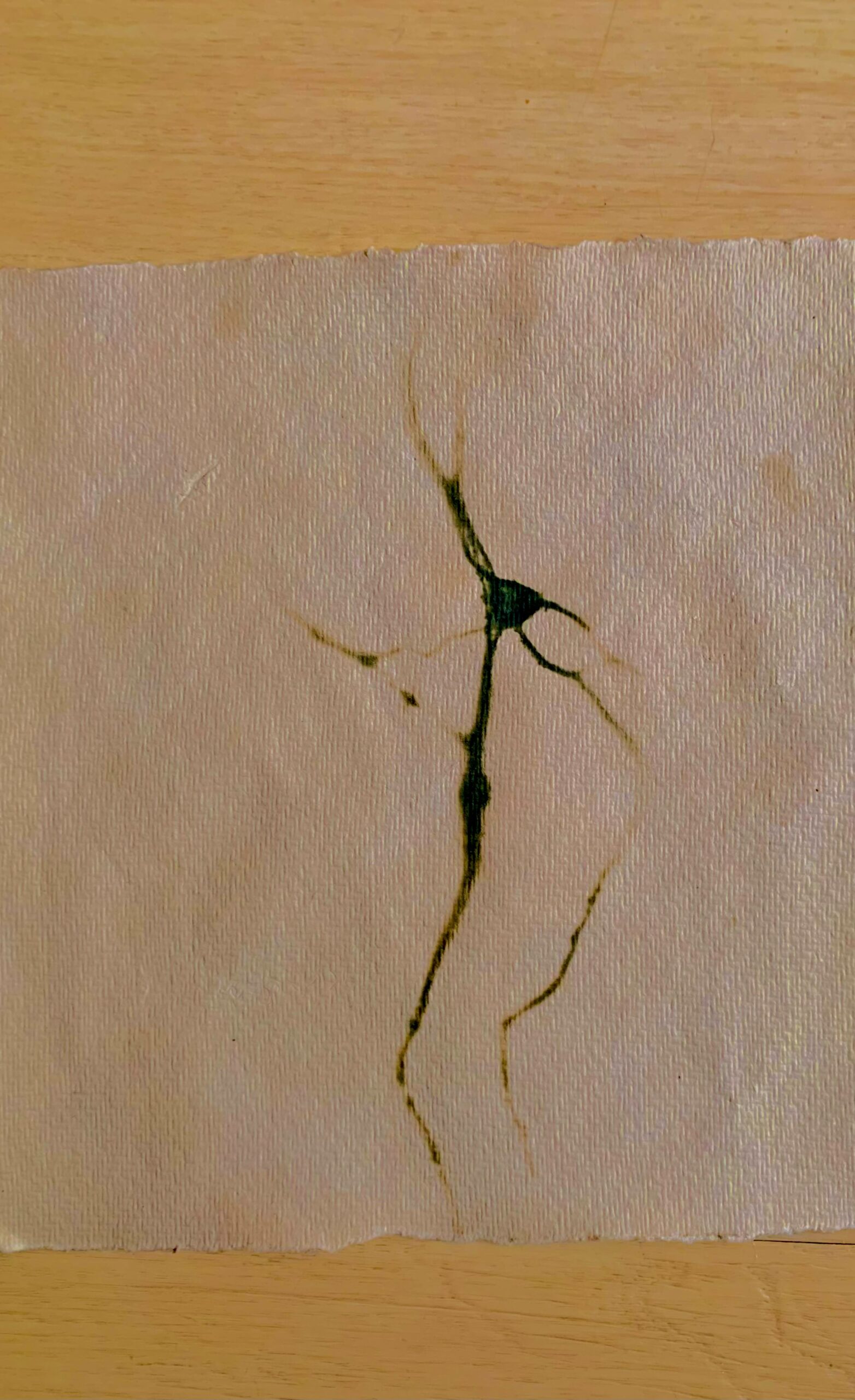
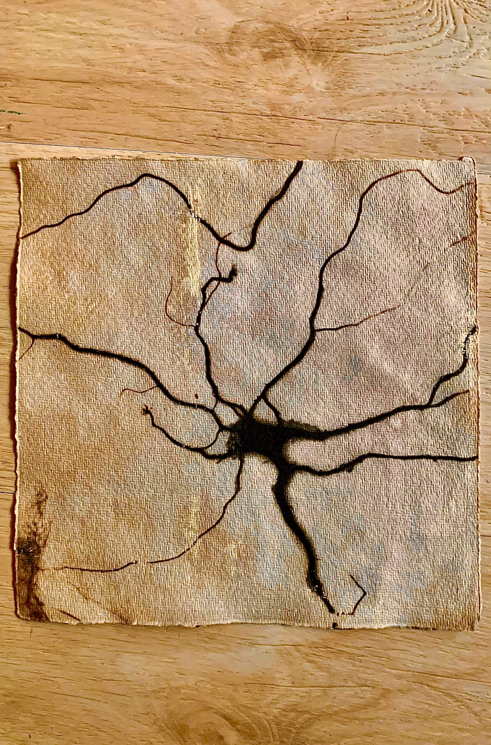
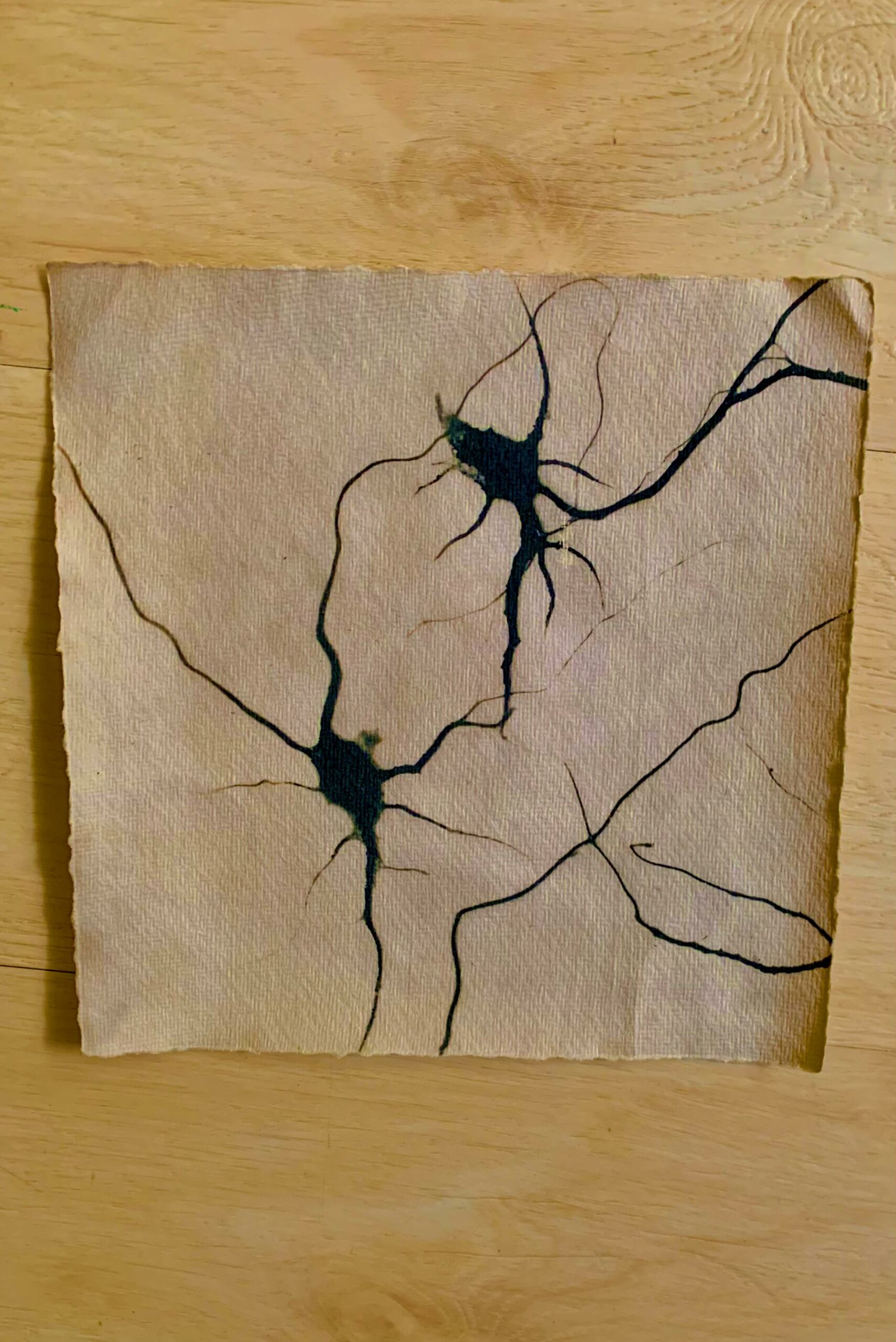
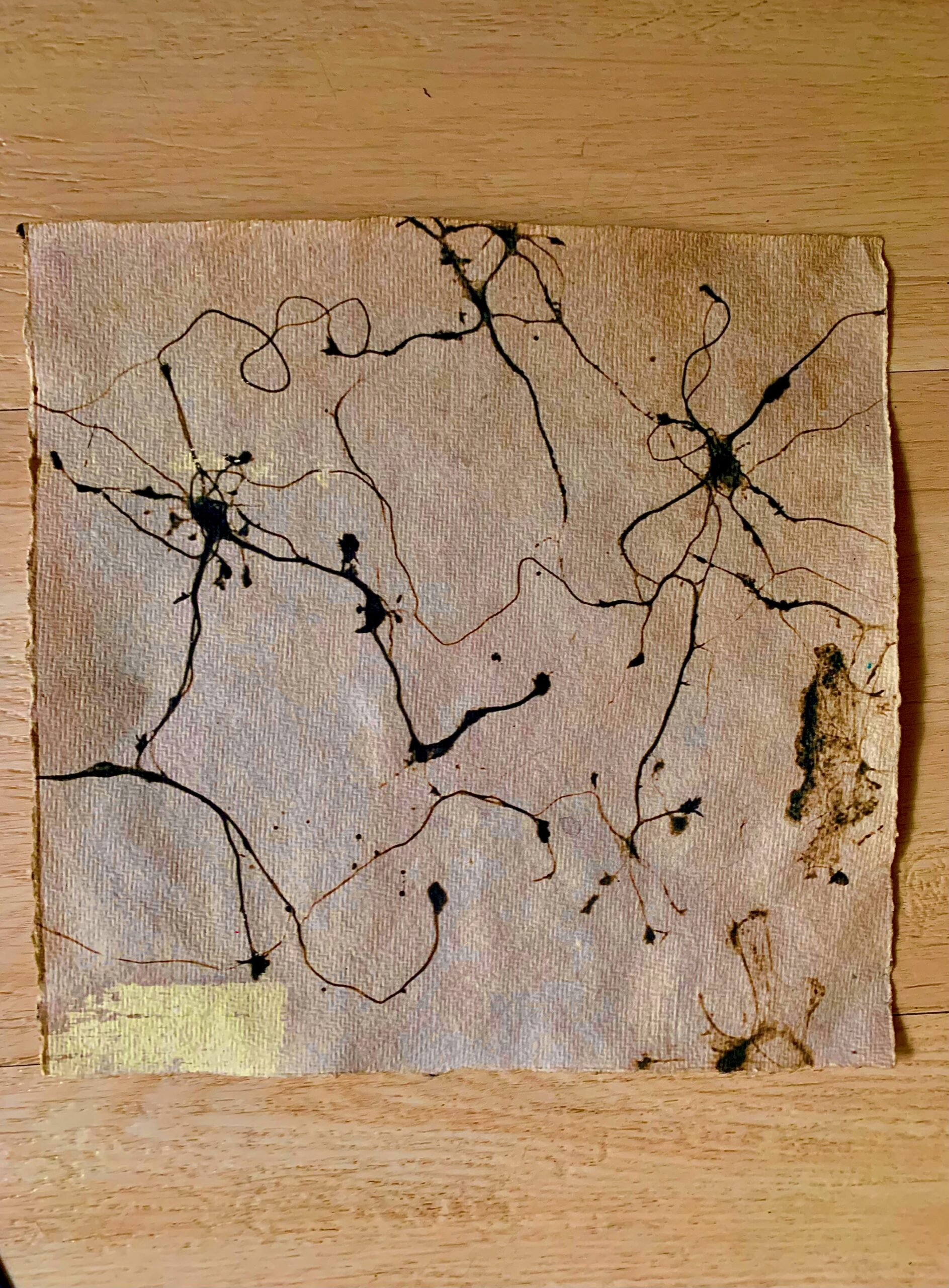
Embryonic rat hippocampal structures were dissected and dissociated to individual neuronal cells which were then allowed to reconnect on a glass coverslip, proliferate, and reform a network of neurons again. These neurons were tracked over time to study their development in culture and were visualized with fluorescent labels these cells. This neuroscientific technique is used to create an in vitro model to study and visualize the subcellular localization and trafficking of neuronal and synaptic proteins, to ultimately understand the molecular mechanisms underlying the development of synapse formation and function.
The high resolution dual labelled fluorescent images were processed as cyanotypes using natural dyes, herbs, and natural UV light exposure. Though this process we have returned the visualisation of these beautiful microscopic cells that form the smallest functional units of our being, into a natural photographic method.
In doing so, the technique brings out new nuances of the natural characteristics of the fluidity of connection of individual brain cells. Some of the natural qualities of these networks harken to the natural patterns of interconnecting fungi or the networks of tree roots. We pair these natural connection designs of neurons in contrast to the grain of wood types and ultimately offer a new perspective on patterns of our natural biology.


Microscopic Connections in Natural Light
Work In Progress with Charlotte Thömmes
Cyanotype prints with natural pigments and UV light.
Details of Individual Pieces
30cm x 40cm
Microscope images, Cyanotype prints with natural pigments and UV light.
Work In Progress
Artists: Tina Ghelani Charlotte Thömmes
Gallery